
Introduction
Babesiosis is a disease caused by protozoal parasites that infect erythrocytes and cause anemia. Babesia species are tick-borne apicomplexan parasites that infect a variety of domestic and wild animals including mammals, marsupials and birds, causing moderate to severe and fatal disease. Human babesiosis is transmitted via ticks from animal reservoir hosts. None of the Babesia species that infect dogs have been found to be zoonotic and infect humans.
Babesiosis has a worldwide distribution and global importance. Dogs are among the animal species in which babesiosis is most common. Hemolytic anemia with erythrocyte destruction and a systemic inflammatory response account for most of the clinical signs observed in Canine Babesiosis.
Canine babesiosis
The initial record of Canine Babesiosis was made in Italy in 1895, not long after the first description of babesiosis in cattle by Victor Babes in Romania (1888). The species causing Babesia infection in dogs was identified in the past based on the morphologic appearance of the parasite.
All large merozoite forms of Canine Babesia (2.5–5.0 μm) were designated Babesia canis, while the small merozoite forms (1.0–2.5 μm) were considered as belonging to Babesia gibsoni. Advances in research and development of molecular methods have demonstrated that additional piroplasmid species infect dogs and cause different pathologies. Babesia rossi, B. canis and B. vogeli, comprising the large form of canine Babesia species, were previously considered as subspecies because they possess identical morphologies on blood smear microscopy. They do differ in the severity of clinical manifestations, genetic characteristics, tick vectors, and geographic distributions therefore are currently classified into separate species. A yet unclassified large Babesia sp. (coco), most closely related to B. bigemina, was found to infect immunocompromised dogs in North America. A Babesia odocoilei-like parasite was reported to have caused canine babesiosis in three dogs from Japan.
The small Babesia spp. that infect dogs include B. gibsoni, B. conradae (originally found in California), B. vulpes (previously termed B. microti-like; Theileria annae) and B. negevi.
The geographical distribution of the causative agents and consequently the occurrence of babesiosis are largely dependent on the habitat of their tick vector species. The exception is B. gibsoni for which evidence for dog-to-dog transmission indicates that infection can be transmitted independent of vector tick infestation. Babesia vogeli and B. gibsoni have wide distributions in both the Old and New World continents, whereas B. rossi has to-date been restricted to sub-Saharan Africa. Babesia canis has mostly been reported from Europe and parts of Asia (Table 1).
Babesia species are transmitted to canine hosts through tick bites. While the infected ticks feed on the dog, Babesia sporozoites are injected with saliva into the host’s skin. The parasites invade the red blood cells, and form ring-shaped trophozoites.
The parasite replicates and forms merozoites seen as pairs of attached pear-shaped inclusion bodies in the erythrocytes in the large Babesia species. Merozoites can divide further to form eight or more parasites in the same erythrocyte and eventually break the red cell escaping into the blood system to invade more erythrocytes (Figure 2).
Ticks feeding on infected blood take up parasites and sexual parasite development of Babesia takes place in the tick gut and is followed by sporogony in its tissues. Parasites reach the tick salivary glands or it’s oocytes from which transmission occurs.
Babesia spp. are transmitted transstadially from one stage in the tick life cycle to another, and also transovarially in some Babesia spp. through the tick eggs. The transmission of babesiae occurs through the bite of a vector tick. But in addition to that, B. gibsoni infection has also been demonstrated to be transmitted via blood transfusion, bite wounds and transplacentally.
Clinical findings in canine babesiosis
It is important to note that the clinical findings in infected dogs vary based on the Babesia species causing the infection and the host’s susceptibility. In general, hemolytic anemia and the systemic inflammatory response syndrome leading to multiple-organ dysfunction syndrome are responsible for most of the clinical signs observed in canine babesiosis. Hemolysis may result in hemoglobinemia, hemoglobinuria, bilirubinemia and bilirubinuria.
Thrombocytopenia is a consistent hematological finding in babesiosis and may be caused by immune mechanisms, splenic sequestration or coagulatory consumption of platelets from hemolytic or vascular injury. Tissue hypoxia is found in severe cases of canine babesiosis. It is caused by anemia, hypotensive shock, vascular stasis by sludging of erythrocytes, excessive endogenous production of carbon dioxide, and consumption of hemoglobin by the parasite.
The central nervous system, kidney, and muscle are affected by hypoxia. Tissue hypoxia, hypertensive shock, multiple organ dysfunction and potential mortality have been documented mostly in B. rossi and B. canis infections. Severe disease with B. vogeli infection is found in young pups and immunocompromised adult dogs, such as dogs with hyperadrenocorticism or treated with immunosuppressive therapy.
The spleen has an important role in controlling babesiosis. Experimentally infected splenectomized dogs rapidly develop parasitemia and clinical disease. Splenectomy has also been associated with fatal outcomes in both human and canine babesiosis.
Diagnosis of babesiosis
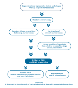
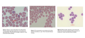
Detection of Babesia in stained blood smears of dogs has been the standard diagnostic technique for canine babesiosis for many years. It is reliable when a moderate to high parasitemia is present. However, in some cases, disease can be present with a low parasitemia which is not readily detected by blood smear microscopy. A direct correlation between the level of Babesia parasitemia and the magnitude of clinical signs is not always found. A fresh smear is recommended for the accurate diagnosis of infection. Microscopy of stained blood smears may show pear-shaped merozoites or ring shaped trophozoites (Figure 1, Figure 2). The small-form Babesia spp. demonstrate smaller shapes without the characteristic pear-shaped architecture (Figure 3). Erythrophagocytosis with infected erythrocytes may be found in blood smears from infected dogs.
Serology is indicative of exposure and not necessarily of a current infection, and therefore may be negative in early infection. There is also serological cross-reactivity between the Babesia species that infect dogs and as a result this test is usually not species-specific.
Molecular diagnostic assays such as PCR are extremely helpful in canine babesiosis. A positive result is indicative of infection, and PCR is especially useful in cases of low parasitemia including suspected carrier dogs or chronically infected animals as well as for speciation of the Babesia species- causing infection. It is also indicated for testing canine blood donors and verifying that they are not infected and will not transmit babesiosis to transfusion recipients. Various PCR techniques are available commercially in specialized laboratories.
The PCRun technique produced by Biogal offers the unique availability to perform PCR in the veterinary hospital or clinic and have results within a short time while the dog and its owners are still waiting for the results of more tests. Biogal offers three PCRun assays for canine babesiosis, the Canine Babesia canis/vogeli molecular detection test, Canine Babesia gibsoni molecular detection test and the newly launched Canine Babesia spp molecular detection test.
Co-infection
Because co-infection is common in canine babesiosis cases, clinicians must be aware that patients with a confirmed diagnosis of babesiosis may be infected with additional tick-borne pathogens that may necessitate a different mode of treatment.
Differential Diagnosis
Babesiosis should be ruled out in cases of IMHA without a clear reason. In addition, babesiosis can in some cases be accompanied by non-regenerative anemia, and it should not be ruled out in these cases.
Treatment of Canine Babesiosis
Several drugs are used to treat canine babesiosis (Table 2). Large Babesia spp. are commonly treated with imidocarb dipropionate with good clinical response while small Babesia spp. appear to be more difficult to treat and resistant to the conventional drugs that are effective against the large babesial species.
Diminazene aceturate used for treatment of both large and small babesial species infections should be used cautiously as it has a relatively small dose safety margin with a large inter-individual pharmacokinetic variation. Babesia gibsoni infection and also infection with other small form Babesia species is often resistant to imidocarb dipropionate and diminazene aceturate. An alternative therapy with the combination of the anti-malarial atovaquone and the macrolide antibiotic azithromycin is recommended for this infection.
However, complete clinical and parasitological cure are not commonly achieved in dogs treated for small Babesia species infections and clinical relapses may occur.
Medical management of infection may require, in addition to the specific anti-protozoal treatment, supportive treatments including blood transfusions, intravenous fluids, and anti-inflammatory drugs. Babesia gibsoni resistance to atovaquone is now widespread in Japan as well as several other countries and alternative treatment protocols with other drug combinations are available to treat this condition.
There is a commercial molecular assay available to detect resistant B. gibsoni strains that have a resistance mutation in the parasite’s Cytochrome b gene.
Prevention of Canine Babesiosis
Prevention of canine babesiosis relies mostly on the avoidance of infectious tick bites. Topical and environmental acaricidal treatments are aimed at reducing the exposure to vector ticks and pathogen transmission to the dog. Babesiosis should be suspected in cases of hemolytic anemia and clinical findings associated with a hemolytic process. Co-infection with other pathogens should be investigated and managed medically if present.
A vaccine against B. canis infection is commercially available in some countries in Europe. The vaccine contains inactivated B. canis soluble antigens obtained from culture medium and is adjuvanted with saponin.
Further reading on canine babesiosis
- Baneth G. Antiprotozoal treatment of canine babesiosis. Vet Parasitol. 2018;254:58-63.
- Baneth G, Florin-Christensen M, Cardoso L, Schnittger L. Reclassification of Theileria annae as Babesia vulpes sp. nov. Parasit Vectors. 2015;8:207.
- Baneth G, Nachum-Biala Y, Birkenheuer AJ, Schreeg ME, Prince H, Florin-Christensen M, Schnittger L, Aroch I. A new piroplasmid species infecting dogs: morphological and molecular characterization and pathogeny of Babesia negevi n. sp. Parasit Vectors. 2020;13:130.
- Checa R, Montoya A, Ortega N, González-Fraga JL, Bartolomé A, Gálvez R, Marino V, Miró G. Efficacy, safety and tolerance of imidocarb dipropionate versus atovaquone or buparvaquone plus azithromycin used to treat sick dogs naturally infected with the Babesia microti-like piroplasm. Parasit Vectors. 2017;10(1):145.
- Criado-Fornelio, A., Gonzalez-del-Rio, M.A., Buling-Sarana et al., The “expanding universe” of piroplasms. Vet Parasitol 2004; 119: 337-345.
- Dear JD, Birkenheuer A. Babesia in North America: An Update. Vet Clin North Am Small Anim Pract. 2022;52:1193-1209.
- Irwin, P.J. Canine babesiosis: from molecular taxonomy to control. Parasit Vectors 2009; 2 Suppl 1, S4.
- Lin EC, Chueh LL, Lin CN, Hsieh LE, Su BL. The therapeutic efficacy of two antibabesial strategies against Babesia gibsoni. Vet Parasitol. 2012;186:159-64.
- Liu M, Igarashi I, Xuan X. Babesia gibsoni. Trends Parasitol. 2022;38:815-816.
- Plumb DC, 2016, Plumb’s Veterinary Drug Handbook, 8th edition. Wiley-Blackwell.
- Solano-Gallego, L., Baneth, G., Babesiosis in dogs and cats-Expanding parasitological and clinical spectra. Vet Parasitol 2011; 181: 48-60.
- Solano-Gallego L, Sainz Á, Roura X, Estrada-Peña A, Miró G. A review of canine babesiosis: the European perspective. Parasit Vectors. 2016;9:336.
- Yamasaki M, Nukada Y, Ito M, Uchida N, Iguchi A, Inokuma H. Three cases of canine babesiosis caused by Babesia odocoilei-like parasites in Japan. Parasitol Int. 2021;84:102384.